
Module 5 Revision: CARE AND PREPARATION OF THE DECEASED
Learning Outcome 1:
Anatomy & Physiology - Lymphatic System
Lymphatic system
The lymphatic system is a network of delicate tubes throughout the body that consists of lymph vessels and lymph nodes. Its main function is to drain fluid (called lymph) that has leaked from the blood vessels into the tissues and empty it back into the bloodstream via the lymph nodes.
The lymphatic system helps to protect us from infection and disease and is part of the body’s immune system. Lymph fluid passes through lymph nodes (filters) which are connected by a network of lymph vessels. The lymph nodes are found throughout the whole body.
The main roles of the lymphatic system include:
-
managing the fluid levels in the body
-
reacting to infection
-
removing cell waste that otherwise would result in disease
-
absorbing some of the fats in our diet from the intestine.
The lymph nodes and other lymphatic structures like the spleen hold special white blood cells called lymphocytes. These can rapidly multiply and release antibodies in response to bacteria and viruses.
How is lymph formed?
Lymph is formed from plasma (the liquid part of blood without the cells). The plasma carries the nutrients needed to ‘feed’ cells all around the body. To reach the cells, plasma must ooze out of capillaries (the smallest blood vessels) into the tissues. Most of this plasma then drains back into the capillaries, but some of it gets ‘left behind’ in the tissues. The fluid left in the tissues drains instead into tiny lymph vessels, where it becomes known as lymph.

Lymph fluid (green) draining from the tissues that is then filtered via the lymph nodes before eventually returning back to the blood.
Lymphatic vessels
The lymphatic vessels are found everywhere in our body. Generally, more active areas of the body have more of them and are comprised of the following:
-
The smaller lymphatic vessels, which take up the fluids from the tissue, are called lymph capillaries.
-
The larger lymphatic vessels have muscles in their walls which helps them gently and slowly pulsate. These larger lymphatic vessels also have valves that stop the lymph flowing back the wrong way.
-
These larger lymphatic vessels then join up (like streams flowing into a river) uand carry the lymph back into the blood, draining into the large veins close to the heart.
-
As the lymph vessels take the lymph back to the blood, they go via the lymph nodes (there are about 700 of these in total in the body ), which are found in our arm pit and groin as well as many other areas of the body such as the mouth, throat and intestines. These work as sieves that filter out (remove) organisms such as bacteria. Like the spleen they are also a home to many lymphocytes that are important for the immune response.
Unlike blood, which is pumped by the heart, lymph is not actively pumped around the system. Instead it is pushed along when the lymph vessels are squeezed by our muscles and or by gravity if the vessel is above the heart.
Spleen overview
The spleen is located in the abdominal (tummy) area on the left side, just under the diaphragm. It is the largest of our lymphatic organs.
The spleen does many things as it filters and monitors our blood. It contains and stores a range of cells, including white blood cells (Lymphocytes) which are important for our body’s defence and can be mobilised at short notice.
As well as removing microbes, the spleen also destroys old or damaged red blood cells. It can also help in increasing blood volume quickly if a person loses a lot of blood.
Lymph nodes - location and structure
As previously discussed lymph nodes are found at various points along the lymph vessels. They are shaped like a bean and usually less than 1 cm long. There are groups of lymph nodes around the body (except for in the brain and spinal cord), for example:
-
In the neck (cervical nodes)
-
Under the arm (axillary nodes)
-
In the groin (inguinal nodes)
-
In the centre of the chest between the lungs (mediastinal nodes)
-
In the abdomen (mesenteric nodes)

Location of the main groups of lymph nodes around the body

This image above shows and individual lymph node and the flow of lymph fluid being filtered through the sinus cavities - filtered lymph fluid eventually returns to the blood stream
Spleen location and functions
-
Store lymphocytes (white blood cells) that can be used to aid our immune systems in times of need.
-
Filter and remove old damaged red blood cells from the circulation.
-
Act as a blood reservoir in times of shock and haemorrhage.
-
Preserves iron when old red cells have been broken down (that can be used to make new haemoglobin).
The spleen contains two types of tissues with different functions: white pulp and red pulp:
Red pulp: Contains cavities filled with blood.
White pulp: Mostly consists of immune cells (white blood cells).
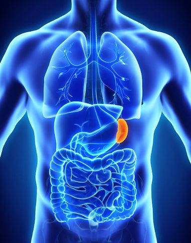
Location of the spleen – its acts like a giant lymph node – but instead of filtering lymph fluid it filters the blood
Tonsils and adenoids
The tonsils and adenoids are part of the immune system. They are similar to the lymph nodes found throughout the rest of your body.
The tonsils are located in the back of your throat and are the two round lumps of tissue you see when you open your mouth wide. You cannot easily see the adenoids as they are found in the upper part of the nasal cavity.


Images above: location of tonsils and adenoids - Tonsillitis is inflammation of the tonsils, typically of rapid onset. Symptoms may include sore throat, fever, enlargement of the tonsils, trouble swallowing, and large lymph nodes around the neck.
The bone marrow, spleen and lymphatic system: immune system
Bone marrow is a semi-solid tissue which may be found within the spongy portions of bones. The major function of bone marrow is to generate blood cells:
-
White blood cells: fight infections
-
Red blood cells: carry oxygen
-
Platelets: help blood to clot

The immune system works within our body to keep us free of infection and disease. It is made up of 3 layers of protection:
-
Physical barriers
-
Immune cells
-
Proteins
Physical barriers: The first way the body is protected against infection is a physical barrier made up of the mucosa (sometimes called mucous membranes) – the soft, moist lining in certain areas of the body, such as the eye, nose, mouth and bowel that are also protected by fluids that wash the mucosa, such as tears, saliva and secretions from the mucosa.
Immune cells: The next line of defence if an organism gets past the physical barrier comes from immune cells - these invlude the lymphocytes.
Proteins: The cells of the immune system work through a number of proteins called antibodies (they are also known as ‘immunoglobulins’) that stick to other proteins called ‘antigens’ on the surface of organisms (such as bacteria and viruses). They directly stop them working, and can break open bacteria or neutralise naturally occurring poisons.
Self Assessment Questions
Q. Name 3 functions of the spleen
Q. Briefly describe the role of bone marrow in relation to the immune system and the 3 cell types it produces.
Q. Draw and label a lymph node - what is the main function of a lymph node?
Q. What is the function of bone marrow?
Q. Explain the main roles of the lymphatic system
Q. What are "lymphocytes"?
Q. The immune system is made up of various organs and tissue (lymphatic system, spleen and bone marrow) and provides 3 layers of protection – what are these 3 layers called?
Q. Where are the adenoids located?
Q. How is ‘Lymph fluid’ formed?
The RF licenses purchased for the above images allows their free use throughout this site