
Module 5 Revision: CARE AND PREPARATION OF THE DECEASED
Learning Outcome 1:
Anatomy & Physiology - Musculoskeletal System
1. Overview of this online learning:
The musculoskeletal systems primary functions include:
-
Supporting the body
-
Allowing motion
-
Protecting vital organs
The musculoskeletal system is made up of:
-
Bones (the skeleton)
-
Muscles
-
Cartilage
-
Tendons
-
Ligaments
-
Joints
-
Connective tissue (that support and bind tissues and organs together).
The skeleton also serves:
-
Main storage system for calcium and phosphorus.
-
Blood production (bone marrow)
2. Skeletal system introduction
The skeletal system includes all of the bones and joints in the body. Each bone is a complex living organ that is made up of many cells, proteins and minerals.
Bones provide a rigid framework known as the skeleton that support and protect the soft organs of the body.
The skeleton supports the body against the pull of gravity and the large bones of the lower limbs support the trunk when standing.
Aided by muscles and ligaments, the vertebrae support the skull and hold the body upright and also provides attachment points for muscles to allow movements at the joints.
The human skeleton is the internal framework of the human body. It is composed of around 270 bones at birth – this total decrease to around 206 bones by adulthood after some bones get fused together. The bone mass in the skeleton reaches maximum density around age 21.
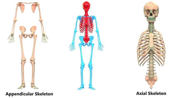
The human skeleton can be divided into the axial skeleton and the appendicular skeleton:
1. The axial skeleton is formed by the:
-
Vertebral column
-
Rib cage
-
Skull
-
Other associated bones
2. The appendicular skeleton, which is attached to the axial skeleton, is formed by the:
-
Shoulder girdle (pectoral girdle)
-
Pelvic girdle
-
Bones of the upper and lower limbs
Subtle differences between sexes in the morphology of the skull, long bones, and pelvis exist. In general, female skeletal elements tend to be smaller and less robust than corresponding male elements within a given population. The human female pelvis is also different from that of males in order to facilitate childbirth.
Functions of the Skeletal System
The skeleton provides the framework of the body and it is made up of 206 bones. It is part of the skeletal system, which is composed of:
-
Bones are tissues that undergo calcification, a process where minerals are deposited to harden the bone.
-
Cartilages are thick and rubbery tissues that are found in joints, the ears, the nose, and the ribs.
-
Ligaments are tough, fibrous tissues that connect one bone to another bone.
-
Tendons are soft tissues that connect muscles to bones.
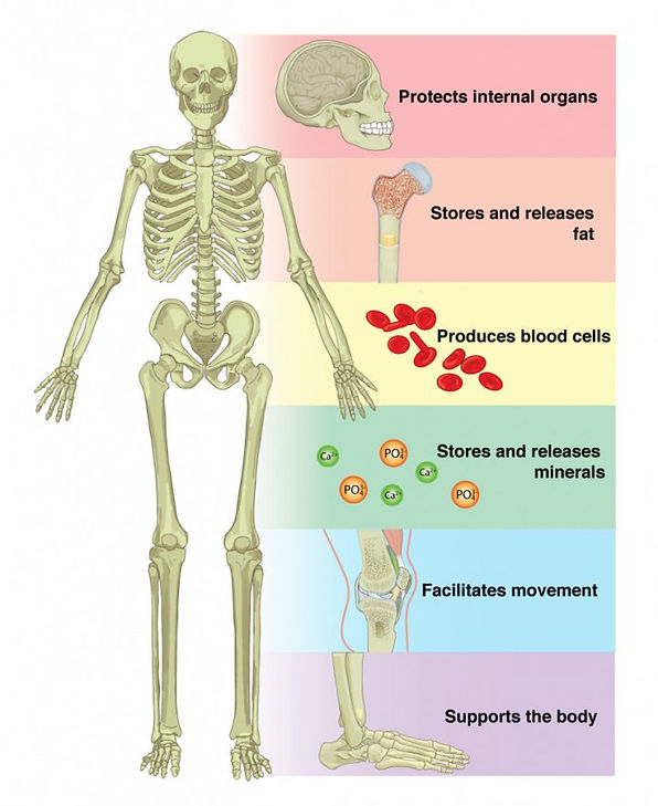
Functions of the skeleton:
1. Shape. The shape of the body is determined by the skeletal framework. Your body shape develops as the skeletal system develops, making you look tall, short, narrow or wide. The size and shape of your hips, hands, feet, and other body parts also follow the shape of your skeleton. These characteristics may be genetically inherited and these are classified as body types such as ectomorphs (thin and tall), mesomorphs (muscular and short), and endomorphs (apple/pear-shaped).
2. Support. Your skeleton supports the body and holds your internal organs in place. The spine or vertebral column lets you stand erect while other bones form hollow spaces to hold organs inside them. Your skull, for example, holds your brain, while your chest cavity contains your heart and lungs. On the other hand, the legs and feet, which are part of the lower appendages, support the body weight.
3. Movement. The bones are attached to other bones and muscles by ligaments and tendons and work together to produce various body movements, making up the musculoskeletal system. Muscles contract and pull bones to produce movements such as walking, running, or lifting. The differences in shapes of the bones play a role in differentiating movements. For example, the small bones in the foot allow you to adapt to walking different terrains, while the small bones of the hands allow you to do detailed, precise movements.
4. Protection. The bony skeleton encases vital organs and protects them from damage. The skull protects the brain, while the spinal column protects the spinal cord and nerves. The ribs and sternum comprise the thorax, which protect your lungs and heart.
5. Blood Cell Production. Long bones contain spongy tissue, which is composed of two types of bone marrow involved in the production of blood cells. Red marrow develops into blood cells. Yellow marrow stores some fat, which develops into red marrow during anaemia or severe depletion of red blood cells.
6. Storage. Bones contain more calcium than any other organ – this mineral is also involved in many important body functions, such as metabolism, blood clotting and nerve transmission. When blood calcium levels decrease below normal, calcium is released from bones so that there will be an adequate supply for metabolic needs. When blood calcium levels are increased, the excess calcium is stored in the bone.
Bone Shapes
The shape of bones reflects their functions:
-
Long bones act as levers to raise and lower.
-
Short bones such as the talus (ankle bone) are useful as bridges
-
Flat bones, including those in the skull, form protective shells.
-
Small rounded, sesamoid bones such as the patella (kneecap) are embedded within tendons.
-
Irregular bones included vertebrae, the ilium (pelvis) and some skull bones, such as the sphenoid.
-
Fused bones, the bones of the face are counted as separate bones, despite being fused naturally. The sacrum is a large, triangular bone at the base of the spine that forms by the fusing of the lower vertebrae between 18 and 30 years of age.
All bones have surface markings and characteristics that make a specific bone unique. There are holes, depressions, smooth facets, lines, projections and other markings. These usually represent passageways for vessels and nerves, points of articulation with other bones or points of attachment for tendons and ligaments.
Skull sutures
In adults, all of the skull bones except the mandible (lower jaw) are locked together by joints known as sutures (see video 1 below). These seams are visible on the surface of the skull as lines between the bones.
From the front, the most prominent skull bones are the frontal bone (cranial bone), which forms the forehead, and the zygomatic bones (facial bone) which give shape to the cheeks and the upper and lower jaw bones.
The back and sides of the cranial vault largely comprise the occipital and parietal bones.
A&P Level 3 (part 2)


Sutures of the Skull | Anatomy Slices

Mucociliary clearance
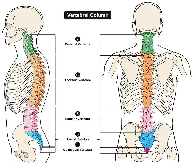
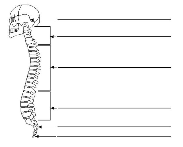
Structure of the Spine
The spine is made up of 33 ring-like bones called vertebrae that are linked by a series of mobile joints. Sandwiched between the vertebrae are springy, shock-absorbing discs with a tough outer layer of cartilage. There 3 main types of Individual vertebrae:
-
x 7 cervical (neck)
-
x 12 thoracic (upper back)
-
x 5 lumbar (lower back)
Also, two regions at the base of the spine consisting of several fused vertebrae:
-
The wedge-shaped sacrum (x 5)
-
The tail-like coccyx (x 4)
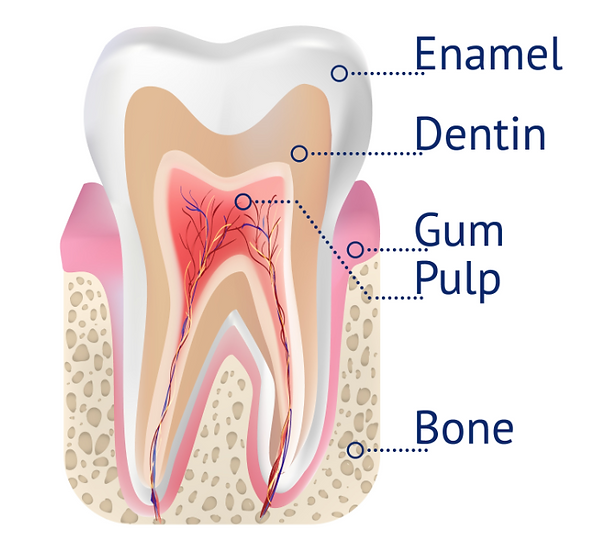
Teeth
The teeth are the hardest substances in the human body. Besides being essential for chewing, the teeth play an important role in speech. Parts of the teeth include:
-
Enamel: The hardest, white outer part of the tooth. Enamel is mostly made of calcium phosphate, a rock-hard mineral.
-
Dentin: A layer underlying the enamel. It is a hard tissue that contains microscopic tubes. When the enamel is damaged, heat or cold can enter the tooth through these paths and cause sensitivity or pain.
-
Pulp: The softer, living inner structure of teeth. Blood vessels and nerves run through the pulp of the teeth.
A normal adult mouth has 32 teeth, which (except for wisdom teeth) have erupted by about age 13:
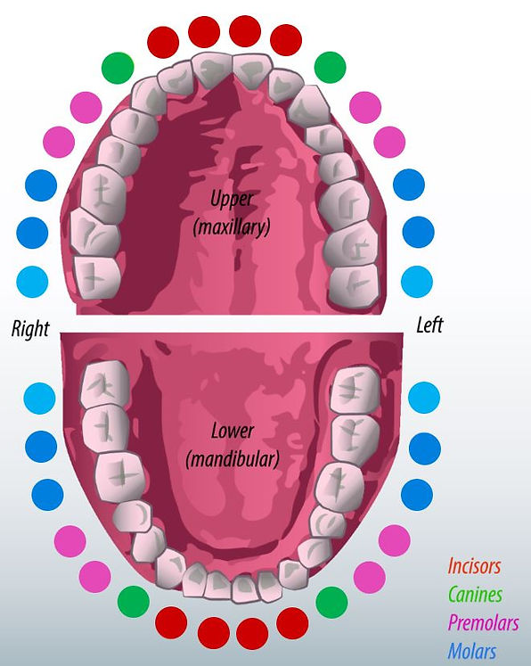
• Incisors (8 total): The middlemost four teeth on the upper and lower jaws.
• Canines (4 total): The pointed teeth just outside the incisors.
• Premolars (8 total): Teeth between the canines and molars.
• Molars (8 total): Flat teeth in the rear of the mouth, best at grinding food.
• Wisdom teeth or third molars (4 total): These teeth erupt at around age 18, but are often surgically removed to prevent displacement of other teeth.
The crown of each tooth projects into the mouth. The root of each tooth descends below the gum line, into the jaw.
Bone Tissue
Although bones vary greatly in size and shape, they have certain structural similarities. Bones have cells embedded in a mineralized (calcium) matrix and collagen fibers. Compact bone forms the shafts of long bones; it also occurs on the outer side of the bone. Spongy bone forms the inner layer.
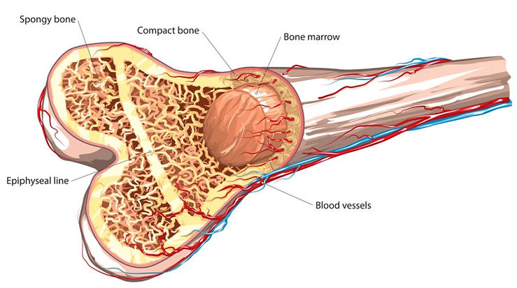
The epiphyseal line the part of the bone that replaces the growth plate in long bones once a person has reached their full adult height.
The growth plate is the portion of the bone that is responsible for a bone’s growth in length during childhood.
Formation of this line takes place over many years. When the growth rate slows down after puberty, the cells in the bone stop the process of replication and all bone growth eventually stops.
After bone growth has stopped the entire growth plate is hardening and turns into bone forming the epiphyseal line.
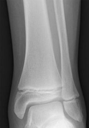
X-ray of tibia (left) and fibula (right) in a child showing two growth plates
Cartilage
Cartilage: is a type of connective tissue that is not as hard and rigid as bone, but is stiffer and less flexible than muscle.
The three types of cartilage found in the body:
-
Hyaline cartilage - is the most common type, found at the junction of long bones, sternum, and ribs. Is a precursor of bone and provides support and strength to different parts of the body. It also provides smooth surfaces, facilitating easy movement of joints.
-
Fibrous cartilage - is found in invertebral discs and joint capsules, and is good at resisting compression.
-
Elastic cartilage - is found in the external ear, epiglottis and larynx and maintains shape and flexibility.
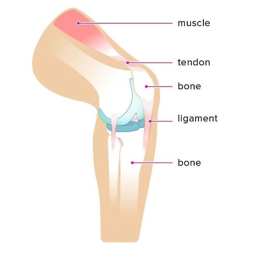
Ligaments and Tendons
Ligaments and tendons are part of the musculoskeletal system, with ligaments attaching bones to bones and tendons muscles to bones. They each serve very important functions to the joints and bones. Ligaments and tendons are made of dense layered collagen fibers, called fibrous connective tissue.
Ligaments
Ligaments serve as connectors, linking the ends of bones together at a joint. The joints allow for the performance of simple and complex motions throughout the body, and ligaments come in a variety of sizes and shapes to support, strengthen and stabilize the joints.
Tendons
Tendons attach muscles to bones. Tendons aid in the movement of bones by transmitting force from the muscle to the bone. Tendons aid in a wide range of motion and act to resist pressures; hence, it is important that they vary in shape and size.
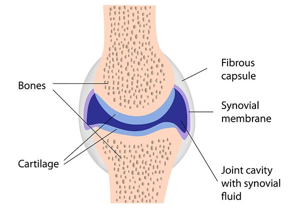
Joints
A joint or articulation is where two bones meet and they connect the bones within your body, bear weight and enable you to move. Joints are classified by their structure or by the way they move (i.e. function).
There are three types of joints: immovable, partly movable, and synovial.
-
Immovable joints, like those connecting the cranial bones, have edges that tightly interlock.
-
Partly movable joints allow some degree of flexibility and usually have cartilage between the bones; example: vertebrae.
-
Synovial joints permit the greatest degree of flexibility and have the ends of bones covered with a connective tissue filled with synovial fluid. Most joints in the body are synovial joints. These versatile lubricated joints allow surfaces in contact to slide over each other easily. example: hip and knee.
3. Muscular system
A muscle is a group of muscle tissues which contract together to produce a force, and consists of specialised cells called muscle fibers that are surrounded by protective tissue and then many more of these fibres bundled together and surrounded in a thick protective tissue. These collective bundles of muscle fibres can collectively produce lots of force.
A muscle uses energy to contract and shorten and are attached to bones (or internal organs and blood vessels), to produce movement. Nearly all movement in the body is the result of muscle contraction.
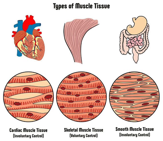
The 3 types of muscle tissue are cardiac, smooth, and skeletal.
1. Cardiac muscle cells are located in the walls of the heart, appear striped, and are under involuntary control.
2. Smooth muscle fibers are located in walls of hollow visceral organs, except the heart, appear irregular spindle-shaped, and are also under involuntary control.
3. Skeletal muscle fibers occur in muscles which are attached to the skeleton. They are striped in appearance and are under voluntary control.
Muscle groups and their actions
Skeletal muscle is the type of muscle that we can see and feel. When a body builder works out to increase muscle mass, skeletal muscle is what is being exercised. Skeletal muscles attach to the skeleton and come in pairs -one muscle to move the bone in one direction and another to move it back the other way. These muscles usually contract voluntarily, meaning that you think about contracting them and your nervous system tells them to do so.
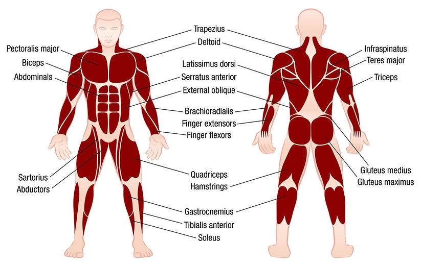
Click here to locate the main muscles mentioned for each of the muscle groups below:
1. The neck
2. The back
3. The shoulder
4. The arm
5. The wrist
6. The hand
7. The abdomen
8. The hip
9. The upper leg and knee
10. The lower leg and foot
4. Self-assessment questions
Q1. Click here and play to quiz to label the skeleton
Q2. Name 3 functions of the skeleton
Q3. What is the difference between tendons and ligaments?
Q4. Muscle tissue can be divided into classes depending on their functionally, or if they are under voluntary or involuntary control; and morphologically (striped or non-striped). By applying these classifications describe the three muscle types.
Q5. What class of muscle would be found in the intestines?
Q6. Name 2 types of cartilage and explain briefly the function of each
Q7. Bones are classified by their shape – give 3 examples
Q8. Name 2 functions of bone joints
Q9. There are three types of joints - name them and give an example for each.
Q10. Briefly describe the general structure of a synovial joint.
Q11. Label the following areas of the vertebral column and state how many bones in each part:
Q12. Briefly describe the main differences between a male and female Skeleton.
Q13. What is an epiphyseal plate?
Q14. What is the function of Synovial fluid?
The RF licenses purchased for these images allows their free use throughout this site