
Module 5 Revision: CARE AND PREPARATION OF THE DECEASED
Learning Outcome 1:
Anatomy & Physiology - Respiratory System
Respiratory system overview
The cells of the human body require a constant stream of oxygen to stay alive. The respiratory system provides oxygen to the body’s cells while removing carbon dioxide, a waste product that can be lethal if allowed to accumulate. There are 3 major parts of the respiratory system: the airway, the lungs, and the muscles of respiration.
-
The airway (upper respiratory tract)
-
The lungs (lower respiratory tract)
-
The muscles of respiration
The airway, which includes the nose, mouth, pharynx, epiglottis, larynx and trachea carries air between from the body's external environment to the lungs.
The lungs act as the functional units of the respiratory system by passing oxygen into the body and carbon dioxide out of the body.
Finally, the muscles of respiration, including the diaphragm and intercostal muscles, work together to act as a pump, pushing air into and out of the lungs during breathing.
The respiratory system also enables speech production.

The airway (upper respiratory tract), which includes the nose, mouth, pharynx, epiglottis, larynx and trachea carries air between from the body's external environment to the lungs.
The lungs act as the functional units of the respiratory system by passing oxygen into the body and carbon dioxide out of the body.
Finally, the muscles of respiration, including the diaphragm and intercostal muscles, work together to act as a pump, pushing air into and out of the lungs during breathing.
Upper Respiratory system
1. Nose and Nasal Cavity
The nose and nasal cavity form the main external opening for the respiratory system and are the first section of the body’s upper respiratory tract through which air moves. The nose is a structure of the face made of cartilage, bone, muscle, and skin that supports and protects the nasal cavity. The nasal cavity is a hollow space within the nose and skull that is lined with hairs and mucus membrane. The function of the nasal cavity is to prepare air before reaching the lungs by:
-
Warming
-
Moisturising
-
Filtering
Hairs and mucus lining the nasal cavity help to trap dust, mould, pollen and other environmental contaminants before they can reach the inner portions of the body.
2. Mouth
The mouth, also known as the oral cavity, is the secondary external opening for the respiratory tract. Most normal breathing takes place through the nasal cavity, but the oral cavity can be used to supplement or replace the nasal cavity’s functions when needed. Because the pathway of air entering the body from the mouth is shorter than the pathway for air entering from the nose, the mouth does not warm and moisturize the air entering the lungs as well as the nose performs this function. The one advantage of breathing through the mouth is that its shorter distance and larger diameter allows more air to quickly enter the body.
3. Pharynx
The pharynx, also known as the throat, is a muscular funnel that extends from the nasal cavity to the oesophagus and larynx.
4. Epiglottis
The epiglottis is a flap of elastic cartilage that acts as a switch between the larynx (airway) and the oesophagus (food passage). Because the pharynx is also used to swallow food, the main function of the epiglottis is to seal off the windpipe during eating, so that food is not accidentally inhaled.

5. Larynx
The larynx, also known as the voice box, is a short tubular section of the airway that connects the pharynx and the trachea (windpipe). Several cartilage ring structures make up the larynx and give it its structure and enable it to produce vocal sounds.
The larynx is about 5 cm (2 in.) long and its outer wall of cartilage forms the area of the front of the neck referred to as the Adam's apple.
6. Trachea
The trachea, or windpipe, is a 5-inch long tube made of cartilage rings and connects the larynx to the bronchi and allows air to pass through the neck and into the thorax. The rings of cartilage making up the trachea allow it to remain open to air at all times.
The main function of the trachea is to provide a clear airway for air to enter and exit the lungs. In addition, the cells lining the trachea produce mucus that traps dust and other contaminants and prevents it from reaching the lungs. Cilia (tiny hair like structures) on the surface of the these cells move the mucus superiorly toward the pharynx where it can be swallowed and digested in the gastrointestinal tract.

Lower Respiratory system
1. Bronchi
A bronchus (or pleural, the bronchi) is either of the two major branches of the trachea that lead to the lungs. The bronchi are the airways that lead from the trachea into the lungs, and then branch into smaller bronchioles. Structurally, the bronchi are made up of cartilage that gives them stability and prevents their collapse. The bronchi function primarily as passageways for air.
The right main bronchus is wider, shorter, and more vertical than the left main bronchus and subdivides into three secondary bronchi (also known as lobar bronchi), which deliver oxygen to the three lobes of the right lung—the upper, middle and lower lobe.
The left main bronchus divides into two secondary bronchi or lobar bronchi, to deliver air to the two lobes of the left lung—the upper and the lower lobe.
Airways such as the bronchi are structures where air exchange does not take place (also known as the conducting portion).

2. Bronchial tree
A bronchial tree (or respiratory tree) is the collective term used for the multiple-branched bronchi. The main function of the bronchi, like other conducting structures, is to provide a passageway for air to move into and out of each lung.
-
Conducting portion of the respiratory system: These structures form a continuous passageway for air to move in and out of the lungs.
-
Respiratory portion of the respiratory system: These thin walled structures allow for gas exchange (Oxygen and Carbon dioxide) to take place.
The bronchi lobes further divide into segmental bronchi, followed by more generations of progressively smaller bronchi. These vessels become progressively smaller and no longer contain cartilage as they divide into bronchioles, terminal bronchioles, respiratory bronchioles, alveolar sacs, and finally into the individual alveoli where the exchange of oxygen and carbon dioxide takes place.

3. Lungs
The lungs are the primary organs of the respiratory system in humans and are located near the backbone on either side of the heart. Their function in the respiratory system is to extract oxygen from the atmosphere and transfer it into the bloodstream, and to release carbon dioxide from the bloodstream into the atmosphere, in a process of gas exchange.
The right lung is bigger than the left, which shares space in the chest with the heart. Each lung is enclosed within a pleural sac which allows the inner and outer walls to slide over each other whilst breathing takes place, without much friction. This sac also divides each lung into sections called lobes. The right lung has three lobes and the left has two.

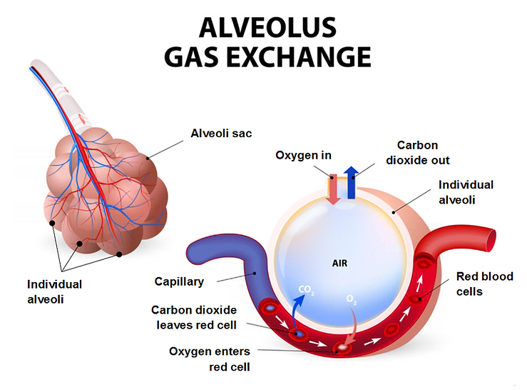
4. Alveoli
The alveoli are located in the respiratory zone of the lungs and are an important part of the respiratory system whose function it is to exchange gas molecules to and from the bloodstream. These tiny, balloon-shaped air structures sit at the very end of the respiratory tree and are arranged in clusters throughout the lungs. The alveoli are only one cell thick, allowing the relatively easy passage of oxygen and carbon dioxide between the alveoli and capillaries.
A typical pair of human lungs contain about 480 million alveoli that provides an approx. 50 -100m squared of surface area. Each alveolus is wrapped in a fine mesh of capillaries covering about 70% of its area.
Muscles of the Respiratory System
The muscles of respiration are those muscles that contribute to inhalation and exhalation, by aiding in the expansion and contraction of the chest cavity. The diaphragm and, to a lesser extent, the intercostal muscles drive respiration during normal breathing.
Maintenance of the elasticity of all muscles used for breathing is crucial to the health of the respiratory system and to maximise its functional capabilities. All muscles that are attached to the human rib cage can aid the breathing action. The speciality of these muscles used for breathing are that they are composed of fatigue resistant muscle fibres, they are controlled by both voluntary and involuntary mechanisms.
1. Diaphragm
The diaphragm is the major muscle responsible for breathing. It is a thin, dome-shaped muscle that separates the abdominal cavity from the chest cavity. During inhalation, the diaphragm contracts, so that its centre moves downward and its edges move upward. This compresses the abdominal cavity, raises the ribs upward and outward and thus expands the thoracic cavity. This expansion draws air into the lungs. When the diaphragm relaxes, elastic recoil of the thoracic wall causes the thoracic cavity to contract, forcing air out of the lungs.
The diaphragm is also involved in non-respiratory functions, helping to expel vomit, faeces, and urine from the body by increasing intra-abdominal pressure, and preventing acid reflux by exerting pressure on the oesophagus.

2. Intercostal muscles
Along with the diaphragm, the intercostal muscles are one of the most important groups of respiratory muscles. These muscles are attached between the ribs and are important in manipulating the width of the rib cage. There are three layers of intercostal muscles. The external intercostal muscles are most important in respiration. These have fibres that are angled downward and forward from rib to rib. The contraction of these fibres raises each rib toward the rib above, with the overall effect of raising the rib cage, assisting in inhalation.

Layers of intercostal muscles in the rib cage that aid breathing
Respiratory support structures
1. Ribs
The ribs help in the expansion and contraction of the thoracic cavity (though that is primarily the function of the diaphragm) and also protect the lungs and heart. The ribs, which are flexible, along with the intercostal muscles, help in this process. Since expansion of the lungs is greater in the lower lobes, the floating ribs enable that process.
2. Pleura
The pleura includes two thin layers of tissue that protect and cushion the lungs. The inner layer (visceral pleura) wraps around the lungs and is stuck so tightly to the lungs that it cannot be peeled off. The outer layer (parietal pleura) lines the inside of the chest wall. The very thin space between the layers is called the pleural cavity. A liquid, called pleural fluid, lubricates the pleural cavity so that the two layers of pleural tissue can slide against each other.

Function
The pleural cavity, with its associated pleurae, aids optimal functioning of the lungs during breathing.
The pleural cavity also contains pleural fluid, which acts as a lubricant and allows the pleurae to slide effortlessly against each other during respiratory movements.
Self assessment questions
Q. What are the 3 major parts of the respiratory system?
Q. Name the areas of the upper respiratory tract.
Q. Name the areas of the lower respiratory tract.
Q. What is the name of the balloon-shaped air structures sit at the very end of the respiratory tree, involved in gaseous exchange?
Q. What is the function of the epiglottis during respiration?
Q. What is the name of the muscles (layers) between the ribs which are important during respiration?
Q. Where do you find the vocal cords?
Q. What is the name of the structure which separates the chest cavity from the abdominal cavity and assists the intercostal muscles during respiration / breathing?
Q. How many lobes are there in the left lung and how many lobes in the right lung?
Q. Each lung is covered with two membranous layers separated by a lubricating fluid. What are they collectively called?
The RF licenses purchased for these images allows their free use throughout this site